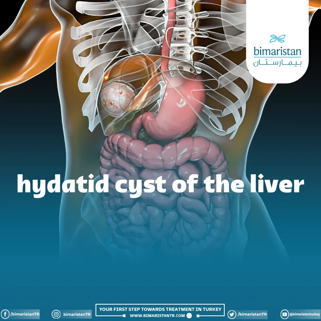
Liver hydatid cyst is a parasitic disease caused by the formation of cysts in the liver. Hydatid cyst treatment involves surgical removal in Turkey.
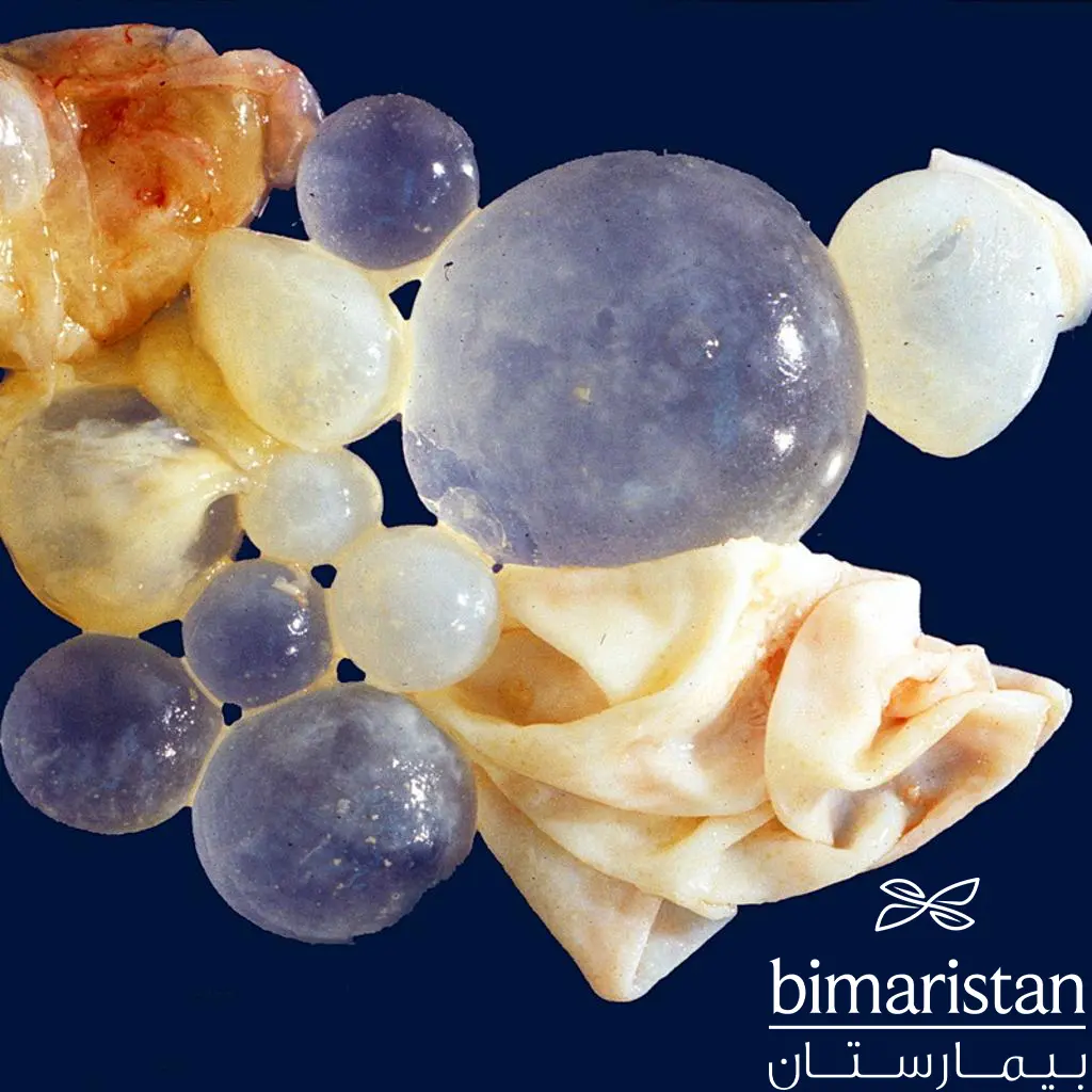
Infection with the Echinococcus granulosus worm (the hydatid cyst parasite) leads to a hydatid disease, often a collection of fluid-filled cysts in the lungs and liver. These cysts are filled with fluid and contain daughter cysts, forming hydatid sand within the fluid when they detach from the cyst. Hydatid cyst in the liver is characterized by slow growth.
Learn about liver hydatid cyst, their causes, and all the details related to their treatment and surgical removal in Turkey.
What is liver hydatid cyst?
Cystic hydatid disease is caused by the Echinococcus granulosus parasite, which affects humans due to ingesting food contaminated with eggs. The eggs hatch inside the human body and go to several organs, including the liver, where hydatid cysts mostly form.
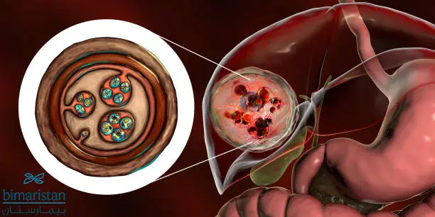
The presence of the hydatid cyst inside the liver with daughter cysts and fluids, along with the surrounding membrane, is noted. The liver is considered the most susceptible organ to infection at 63%, followed by the lungs at 25%, then muscles at 5%, bones at 3%, kidneys at 2%, brain at 1%, and spleen at 1%. The uterus infection is considered a rare case.
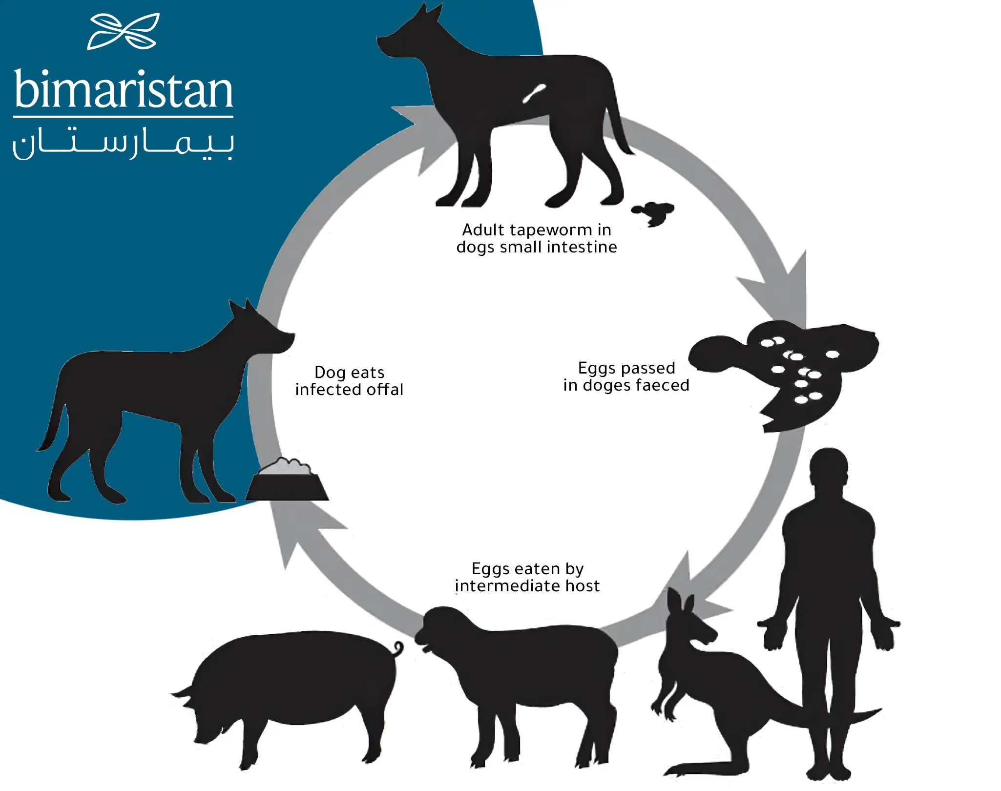
The life cycle of the Echinococcus granulosus worm
- The adult worm is present in the intestines of the definitive host (the dogs).
- The eggs of the Echinococcus worm are excreted with the feces of the definitive host, and they become infectious immediately by consuming vegetables contaminated with these eggs.
- The larva enters the intestines of an intermediate host (such as humans) and hatches to give rise to a spiky larva that penetrates the intestines of humans.
- The larva (hydatid parasite) migrates through the blood and forms hydatid cysts in one of the body’s organs, forming hydatid cysts in the liver (usually affecting the liver and lungs).
When a definitive host consumes the liver of an intermediate host, the primary larva enters the intestines and attaches to the digestive system, then develops into an adult worm in their intestines. However, in the definitive host, it does not lead to the formation of a hydatid cyst.
Cystic hydatid disease causes
Consuming food and water contaminated with Echinococcus granulosus eggs leads to infection and the formation of cysts on the liver most of the time. Due to this transmission mode, hydatid disease is not contagious through human contact.
However, if the water inside the hydatid cyst in the liver leaks along with daughter cysts, it can be implanted anywhere it reaches and lead to the formation of new cysts.
Liver hydatid cyst symptoms
Most cysts do not cause symptoms because hydatid cysts grow slowly and do not lead to rapid onset of symptoms. However, if the cyst grows, symptoms appear due to pressure on adjacent organs.
Therefore, when it is small, it may cause vague symptoms such as fever. A hydatid cyst in the liver may be painful and lead to a feeling of bloating in the abdomen. It may also lead to increased pressure in the portal vein if it is close to it, leading to its blockage.
However, when the cyst becomes larger, it leads to symptoms due to pressure on adjacent structures. For example, symptoms of hydatid disease may lead to obstructive jaundice and abdominal pain, with tearing at the level of adjacent bile ducts. The classic triad of obstructive jaundice, biliary colic pain, jaundice, and biliary itching has also been observed.
But it is also possible for cysts to be present in the lungs as well as in the liver or alone, leading to additional symptoms such as coughing, shortness of breath, side pain, and bloody sputum. Cysts can also be present in the brain, leading to headaches, dizziness, and other neurological symptoms depending on the affected area.
Liver hydatid cyst risk
The echinococcal disease risk comes when it ruptures or bursts. In case of mild leakage of cyst contents, it may lead to a mild allergic reaction consisting of redness and itching.
But in case of major rupture or bursting, an allergic reaction occurs with a severe drop in blood pressure that may lead to death.
Hepatic hydatid cyst diagnosis
Physical examination does not show specific signs, but hepatomegaly or respiratory slip signs may be observed. Laboratory tests and radiological imaging are two essential methods for diagnosing liver cysts in Turkey.
Laboratory tests for hepatic hydatid disease
Routine blood tests are not crucial for confirming diagnosis but may show an increase in white blood cell count at the expense of eosinophils. Liver involvement may be reflected by elevated bilirubin and alkaline phosphatase levels, which are supportive tests.
Indirect hemagglutination tests and enzyme-linked immunosorbent assay (ELISA) have a sensitivity of 80% for detecting hydatid disease (about 90% for liver infections), so they are the first to detect the disease.
Casoni test
In the past, parts of the parasite were injected into the skin, which was a test for parasite sensitivity, and the sensitivity of this test was 70%. However, this test has been abandoned due to its low sensitivity and accuracy and because it can lead to severe local allergic reactions.
Radiological tests
Radiological imaging methods used to detect liver hydatid cysts include:
Simple X-rays
In the best-case scenario, chest and abdominal X-rays are not diagnostic and do not reveal cysts. Simple calcifications around the tumor may indicate a hydatid cyst, but it is not definitive. In lung cysts, chest X-rays may sometimes appear normal.
Diagnostic criteria for radiological imaging:
- Multilayered hydatid cyst wall
- Thick hydatid cyst wall in the liver with some calcifications
Ultrasound imaging
Ultrasound imaging may help diagnose the infection if the doctor sees daughter cysts inside the liver hydatid cyst and if they see hydatid sand. However, the usefulness of ultrasound imaging is limited to the experience of the performing radiologist.
Computed tomography (CT) imaging
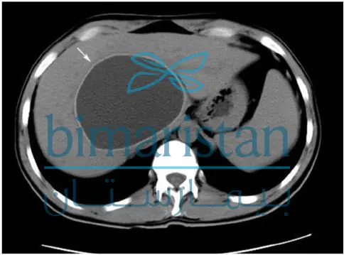
The best procedure for diagnosing liver hydatid cysts, with an accuracy of about 98%, and has the ability to detect daughter cysts inside the main cyst. It is the best for distinguishing between them and amoebic cysts, abscesses in the liver, and liver fibrosis.
We notice the presence of the hydatid cyst in the liver and the fluid inside it on axial CT imaging, and we also see the thick membrane of the hydatid cyst.
Magnetic resonance imaging (MRI)
MRI shows daughter cysts well, but it does not provide any additional benefit over CT imaging.
Endoscopic retrograde cholangiopancreatography (ERCP)
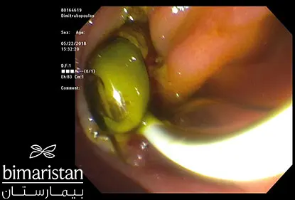
It provides diagnostic and therapeutic benefits as the doctor enters an endoscope into the bile ducts to diagnose and help treat any cysts present in the bile ducts. However, it cannot be them if they are present in the liver or on the external surface of the liver.
Liver hydatid cyst treatment
There are many methods for treating liver cysts, but the most important method is surgical removal of the cyst in Turkey, which ensures a high cure rate. These methods include:
Hydatid cyst medical treatment
Small, single, superficial cysts may respond to antiparasitic drugs, especially albendazole, which kills the parasite and reduces the size of liver cysts.
PAIR technique for liver cystic echinococcosis treatment
This is one of the modern techniques in Turkey, which means puncturing, aspirating, injecting parasiticidal agents, and then reaspirating. This technique is performed using a needle or catheter to puncture the hydatid cyst and draw out its contents, then injecting a parasiticidal chemical and leaving it for a while before aspirating it. The doctor repeats this process until the cyst is completely drained and excised and removed.
Liver hydatid cyst surgery in Turkey
Surgical removal of the hydatid cyst in Turkey is the best option for treating large single or mixed (ruptured or containing a fistula with the bile duct) liver cysts or those with a diameter greater than 10 cm.
Albendazole should be given one week before surgical removal of a liver hydatid cyst in Turkey to reduce the size of liver cysts and reduce the risk of secondary infection or hypersensitivity if fluid leaks into the abdominal cavity. It should also protect the surgical field (cover the liver) with screens moistened with a high-concentration saline solution (e.g., 20%).
The cyst should be excised in one piece, known as surgical enucleation of the hydatid cyst. This is done with great precision to avoid perforating the liver hydatid cyst – Bimaristan Hospital Center in Turkey provides the best doctors in this field. In cases where enucleation of the cyst is complex, the PAIR technique is used to remove it.
Hydatidiform cyst surgical intervention indications
If the liver hydatid cyst is large, single, superficially located on the liver, contains multiple daughter cysts, carries a risk of rupture, is inside the bile ducts, or is pressing on vital organs, it requires surgical removal.
Cysts on other organs, such as the brain, kidneys, and others, also require surgical removal. The surgical procedure is performed by a medical team led by a general surgeon, who may prefer to combine these methods to get rid of the liver hydatid cyst.
Follow-up after surgery
Liver hydatid cyst disease is a recurring parasitic infection, so it is recommended to follow up with imaging procedures such as liver ultrasound or computed tomography for up to three to five years to ensure the body’s safety.
Herbal treatment for hydatid cysts
Many parasitic and bacterial diseases are treated with herbs; some have shown a good effect on parasites. However, research is still ongoing in laboratories for hydatid disease, and some drugs still have bad side effects. Currently, research is underway for herbal treatment for liver hydatid cysts.
Liver hydatid cyst prevention
Since the infection is caused by consuming food contaminated with eggs, prevention is done by following these steps:
- Educating about personal hygiene
- Controlling pet food (for example, stopping feeding sheep offal to dogs)
- Treating pets for intestinal hydatid disease in endemic areas
- Not using sewage water contaminated with hydatid tapeworm for agriculture or drinking
Finally, liver hydatid cyst disease, which arises from the Echinococcus granulosus parasite, leads to the formation of hydatid cysts in the liver, lungs, or other areas. These cysts can cause symptoms due to pressure on adjacent structures or due to cyst rupture, leading to anaphylactic shock that can be fatal. Therefore, it is a disease that requires treatment, and Turkey has the best-specialized centers. It is always preferable to try to prevent the disease in the community.
Sources:
- Centers for Disease Control and Prevention
- Medscape
- UpToDate

