جراحة الديسك المجهرية
جراحة الديسك المجهرية Microdiscectomy Surgery تعتبر المعيار الذهبي للتخلص من آلام الديسك التي يعاني منها كبار السن لذا فهي تجرى بيد أمهر جراحي العصبية في تركيا.

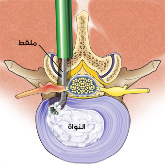
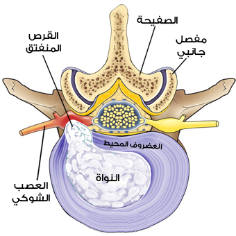
جراحة الديسك المجهرية الدقيقة، هي استئصال الديسك المنفتق مجهرياً، وبذلك يخف الضغط على جذر العصب الشوكي عن طريق إزالة المادة المسببة للألم.
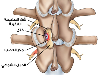
أثناء العملية، قد يتم استئصال جزء صغير من العظم الموجود فوق جذر العصب و / أو مادة القرص الموجودة أسفل جذر العصب.
عادة ما يكون استئصال ديسك الغضروف المنفتق عبر المجهر (والتي تسمى أيضاً إزالة الضغط حول العصب المجهرية) أكثر فعالية في تخفيف آلام الساق (المعروف أيضًا باسم اعتلال الجذور أو عرق النسا) من آلام أسفل الظهر:
بالنسبة لألم الساق، سوف يشعر المرضى عادةً بتخفيف الآلام فورًا بعد استئصال القرص الغضروفي بمساعدة المجهر.
وفي معظم الأحيان يعودون إلى المنزل بعد الجراحة بعد أن تكون آلامهم قد زالت تماماً.
بالنسبة للخدر أو الضعف أو أي أعراض عصبية أخرى في الساق أو القدم، قد يستغرق الأمر أسابيع أو حتى أشهر حتى يلتئم جذر العصب تمامًا ويهدأ أي خدر أو ضعف.
فبالرغم من فائدتها الآنية في مجال علاج الألم إلا أن شفاء العصب قد يستغرق عدة أشهر.
كقاعدة عامة، يعتبر استئصال القرص المجهري عملية جراحية موثوقة نسبيًا للتخفيف الفوري، أو شبه الفوري، من آلام عرق النسا الناتجة من القرص الغضروفي القطني.
عمليات جراحة الديسك طفيفة التوغل
هناك خياران شائعان في العيادة الخارجية لاستئصال القرص الغضروفي القطني:
- جراحة الديسك المجهرية الدقيقة.
- جراحة الديسك بالمنظار (أو عن طريق الجلد).
بالإضافة لوجود خيار ثالث بدون جراحة وهي علاج الديسك بالليزر.
تعتبر ﺟﺮاﺣﺔ اﺳﺘﺌﺼﺎل الديسك المجهرية الدقيقة بشكل عام المعيار الذهبي لإزالة الجزء المنفتق من القرص الذي يضغط على العصب، حيث إن هذا الإجراء له تاريخ طويل والعديد من جراحي العمود الفقري لديهم خبرة واسعة في هذا النهج.
أثناء الجراحة المفتوحة من الناحية الفنية، تستخدم عملية استئصال القرص المجهري تقنيات طفيفة التوغل ويمكن إجراؤها من خلال شق صغير نسبيًا مع الحد الأدنى من تلف الأنسجة أو اضطرابها.
اكتسب بعض الجراحين الآن خبرة كافية في تقنيات التنظير الداخلي أو تقنيات الجراحة عبر المنظار، والتي تتضمن إجراء الجراحة من خلال أنابيب يتم إدخالها في منطقة الجراحة، وليس من خلال شق مفتوح.
عادة ما يتم إجراء استئصال القرص المجهري بواسطة جراح العظام أو جراح الأعصاب.
أسباب التداخل لجراحة الديسك المجهرية (الانزلاق الغضروفي)

بشكل عام إذا كان الألم في الساق الناتج عن الديسك يتحسن، ففي معظم الأحيان سوف يزول الألم بشكل كامل في غضون ستة إلى اثني عشر أسبوعًا من بداية الألم.
طالما أن الألم يمكن تحمله ويمكن للمريض أن يعمل بشكل مناسب، فمن المستحسن عادةً تأجيل الجراحة لفترة قصيرة من الوقت لمعرفة ما إذا كان الألم سيختفي بالعلاج غير الجراحي وحده.
وفي بعض الأحيان من الممكن تجربة العلاج عبر الليزر
ومع ذلك إذا كان ألم الساق شديدًا جداً، فمن المعقول أيضًا التفكير في الجراحة بشكل عاجل.
على سبيل المثال، إذا كان المريض يعاني من ألم شديد على الرغم من استنفاذ جميع طرق العلاج غير الجراحية بحيث يصعب عليه النوم أو الذهاب إلى العمل أو أداء الأنشطة اليومية، فيمكن عندها التفكير في الجراحة.
الأسباب النموذجية للتوصية بإجراء العلاج الجراحي بواسطة المجهر:
- استمرار ألم الساق لمدة ستة أسابيع على الأقل.
- وجود فتق في الديسك في تقرير التصوير بالرنين المغناطيسي أو أي اختبار آخر.
- إذا كان ألم الساق (عرق النسا) هو العرض الرئيسي للمريض، وليس مجرد آلام أسفل الظهر.
- إذا كانت العلاجات غير الجراحية مثل الستيروئيدات عن طريق الفم ومضادات الالتهاب غير الستيروئيدية والعلاج الطبيعي لم تخفف الآلام بشكل كافٍ.
في حال وجود إحدى هذه الحالات تكون نتائج الجراحة أسوء ا إلى حد ما عند مرور ثلاثة إلى ستة أشهر منذ ظهور الأعراض، لذلك ينصح الأطباء عادة الأشخاص بعدم تأجيل الجراحة لفترة طويلة (أكثر من ثلاثة إلى ستة أشهر).
خطوات علاج الديسك نهائياً عبر جراحة الديسك المجهرية في تركيا (الانزلاق الغضروفي)
في معظم الحالات يتم إجراء استئصال القرص المجهري من خلال الظهر، بحيث يستلقي المريض على طاولة العمليات الجراحية ووجهه للأسفل.
يتم استخدام التخدير العام، وعادة ما تستغرق العملية حوالي ساعة إلى ساعتين.
خطوات الجراحة النموذجية:

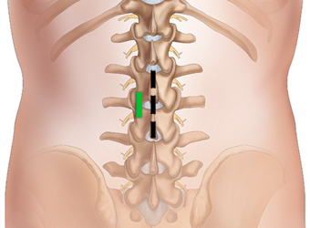
يقوم الدكتور أخصائي المخ والأعصاب وجراحة العمود الفقري بإجراء استئصال القرص المجهري من خلال فتح شقوق لا تتجاوز 5 سم في خط الوسط من أسفل الظهر.
أولاً: يتم رفع عضلات الظهر من قوس الفقرات (الصفيحة) للعمود الفقري وتحريكها جانباً.
نظرًا لأن عضلات الظهر هذه تعمل عموديًا، يتم تثبيتها على الجانب باستخدام المباعد أثناء الجراحة؛ حيث لا يوجد حاجة لقطع العضلات.
يستطيع الجراح بعد ذلك دخول العمود الفقري والوصول للديسك عن طريق إزالة الصفاق الموجود فوق الفقرات (ligamentum flavum).
تسمح النظارات المكبرة أو المجهر الالكتروني للجراح بتصور جذر العصب بشكل واضح.

في بعض الحالات، يتم إزالة جزء صغير من مفصل الوجه الداخلي للفقرة لتسهيل الوصول إلى جذر العصب ولتخفيف أي ضغط أو انضغاط على العصب.
وأحياناً قد يقوم الجراح بعمل فتحة صغيرة في الصفيحة العظمية للفقرة إذا لزم الأمر للوصول إلى موقع الجراحة.
يتم تحريك جذر العصب برفق إلى الجانب.
يستخدم الجراح أدوات صغيرة للذهاب تحت جذر العصب وإزالة زوائد مادة القرص التي تنبثق من الديسك.
تعود العضلات إلى مكانها.
يتم إغلاق الشق الجراحي وتوضع شرائط معقمة فوق الشق للمساعدة في تثبيت الجلد في مكانه للشفاء.
في استئصال الديسك المجهري، تتم إزالة الجزء الصغير فقط من القرص الذي انفتق – أو تسرب من القرص.
حيث يتم ترك غالبية القرص كما هو.
الأهم من ذلك، لأن جميع المفاصل والأربطة والعضلات بقيت سليمة، فلا يغير استئصال القرص المجهري التركيب الميكانيكي للعمود الفقري السفلي للمريض (العمود الفقري القطني).
بعد الجراحة
يمكث المرضى عادةً في المستشفى لبضع ساعات بعد الجراحة قبل خروجهم للعودة إلى المنزل.
اعتمادًا على حالة المريض، قد يوصى بإقامة ليلة واحدة في المستشفى.
بعد العملية، قد يعود المرضى إلى المستوى الطبيعي نسبيًا من الأنشطة بسرعة.
عادة ما يتم تشجيع المرضى على المشي في غضون ساعات قليلة من الجراحة.
سيقدم الجراح تعليمات الرعاية المنزلية، بما في ذلك الأدوية، النشاطات المنزلية، ونوع المتابعة، وغيرها من المعلومات.
المخاطر والمضاعفات بعد عملية الديسك ومعدلات النجاح
تعتبر هذه الجراحة التي يتم إجراؤها على نطاق واسع، أنها تتميز بمعدلات نجاح عالية نسبيًا، خاصة في تخفيف آلام ساق المرضى (عرق النسا).
حيث إنه عادة ما يكون المرضى قادرين على العودة إلى المستوى الطبيعي للنشاط بسرعة إلى حد ما.
معدلات نجاح استئصال القرص المجهري
معدل نجاح جراحة العمود الفقري المجهري مرتفع بشكل عام، حيث أظهرت دراسة طبية واسعة النطاق نتائج جيدة أو ممتازة بشكل عام لـ 90٪ من الأشخاص الذين أجروا العملية.
تشير الدراسات الطبية أيضًا إلى بعض الفوائد للجراحة، عند مقارنتها بالعلاج غير الجراحي، على الرغم من أن الاختلاف يقل بمرور الوقت في بعض الحالات.
حيث وجدت إحدى الدراسات الكبيرة أن الأشخاص الذين خضعوا لجراحة الانزلاق الغضروفي القطني لديهم تحسن أكبر في الأعراض لمدة تصل إلى عامين مقارنة بأولئك الذين لم يخضعوا لعملية جراحية.
تكرار حدوث الانزلاق الغضروفي
تختلف التقديرات، ولكن ما بين 1٪ و20٪ من الأشخاص الذين أجروا استئصال الديسك المجهري سيصابون بفتق قرصي آخر في مرحلة ما.
قد يحدث انفتاق القرص الإضافي مباشرة بعد جراحة الظهر أو بعد عدة سنوات، على الرغم من أنه أكثر شيوعًا في الأشهر الثلاثة الأولى بعد الجراحة.
إذا انفتق الديسك مرة أخرى، فسيكون معدل نجاح العملية الثانية مساوي تقريباً للعملية الأولى ومع ذلك، بعد تكرار المرض، يكون المريض أكثر عرضة لتكرار الإصابة مرة أخرى.
بالنسبة للمرضى الذين يعانون من تكرار الانزلاق الغضروفي المتعدد، قد يوصى بدمج العمود الفقري لمنع تكرار حدوثه مرة أخرى.
تعد إزالة القرص بالكامل ودمج المستوى هو الطريقة الأكثر شيوعًا لضمان عدم حدوث المزيد من الانزلاق في الغضروف.
إذا لم يتم المساس بمفصل الوجه الخلفي وتم استيفاء بعض المعايير الأخرى، فيمكن النظر في استبدال القرص الاصطناعي(الصفائح)، واستبداله مكان الديسك المصاب.
بعد جراحة العمود الفقري المجهري، يوصى ببرنامج تتمارين للتمدد والتقوية للمساعدة في منع تكرار آلام الظهر أو حدوث الديسك.
المخاطر والمضاعفات المحتملة لاستئصال القرص المجهري
كما هو الحال مع أي شكل من أشكال جراحة العمود الفقري، هناك العديد من المخاطر والمضاعفات المرتبطة باستئصال الديسك المجهري.
يحدث تمزق في غشاء الأم الجافية مما يؤدي إلى (تسرب السائل النخاعي) في حوالي 1٪ إلى 7٪ من جراحات استئصال الديسك المجهري.
لا يغير التسريب من نتائج الجراحة، ولكن قد يُطلب من المريض الاستلقاء لمدة يوم إلى يومين بعد الجراحة لحين توقف التسريب.
تشمل المخاطر والمضاعفات الأخرى ما يلي:
- تلف جذر العصب.
- سلس الأمعاء / المثانة.
- نزيف.
- عدوى.
- تراكم محتمل للسوائل في الرئتين قد يؤدي إلى الالتهاب الرئوي.
- تجلط الأوردة العميقة، والذي يحدث عندما تتشكل جلطات دموية في الساق.
- ألم يستمر بعد الجراحة.
انظر متلازمة جراحة الظهر الفاشلة (FBSS): ما هي وكيفية تجنب الألم بعد الجراحة
المضاعفات المذكورة أعلاه ليست شائعة كثيراً لجراحة العمود الفقري المجهري.
