What is a pericardial effusion?
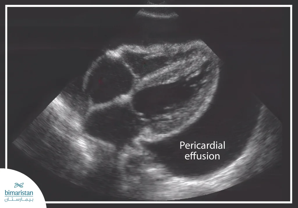
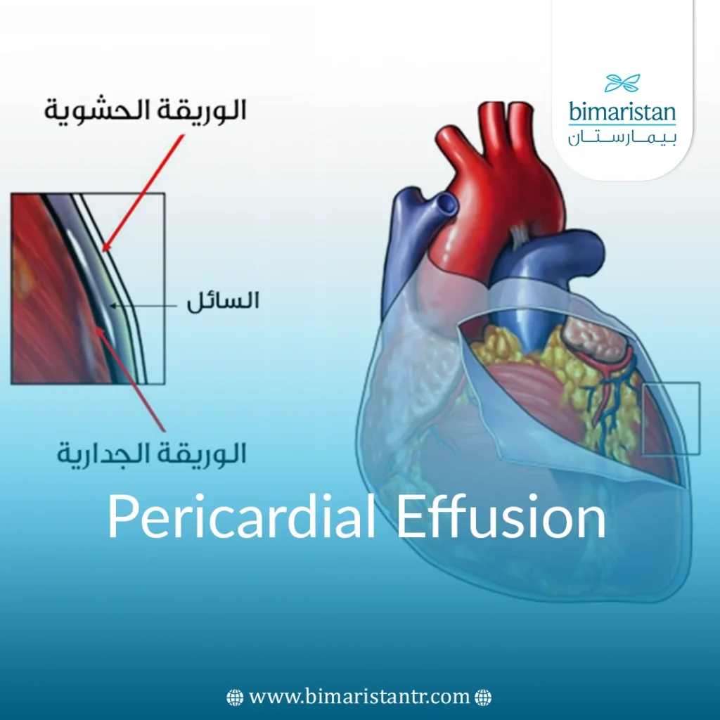
Pericardial effusion is the collection and accumulation of fluid within the pericardium, which surrounds the heart.
The volume of fluid within the pericardial sac may increase due to bleeding or effusion (e.g., due to pericarditis).
The amount of fluid in a pericardial effusion depends on the amount of fluid that has accumulated, typically ranging from 20 to 50 mL. In extreme cases, such as those with tuberculous pericarditis, the volume can increase to as much as two liters.
An effusive pericardium is rare.

What happens if there is an increased accumulation of fluid causing pericardial effusion in the pericardial cavity (heart sac)?
Since the outer layer of the pericardium is not very elastic, there is increased pressure on the heart muscle when an effusion occurs.
This restricts the heart’s activity. It hinders the heart chambers from filling with enough blood.
However, the pressure in the heart chambers continues to rise. As a result, the heart cannot pump enough blood to the body.
Pericardial effusion symptoms
Smaller pericardial effusions are often asymptomatic.
In other cases, the following symptoms occur:
- Pain behind the sternum or tightness (especially when lying down and when breathing)
- General weakness
- Upper abdominal pain with hepatomegaly and congestive cirrhosis (cardiac cirrhosis)
- Tachypnea and shortness of breath
- Low blood pressure
- Tachycardia, palpitations
- Dysphagia (esophageal compression)
- Hoarseness (pressure on the recurrent laryngeal nerve)
- Hiccups (phrenic nerve compression)
- Coughing (compression of the trachea or bronchi)
Depending on the cause, there may be additional symptoms, such as fever in the case of infectious pericarditis.
After analyzing all cases of pericardial effusion, the following signs were found in the case of acute pericardial effusion or cardiac pericardial effusion:
- Low blood pressure
- Tachycardia
- Jugular vein congestion
- Pallor (peripheral vasoconstriction)
- Shortness of breath, tachypnea
- Cardiogenic shock
- Asystole stops
- Sympathetic symptoms: Sweating, insomnia
Possible causes of all pericardial effusions
Pericardial effusion is a common result of pericarditis. Pericarditis, which can lead to pericardial effusion, is caused by:
- Infectious diseases (mostly caused by viruses)
- Rheumatic inflammation (due to autoimmune diseases)
- Malignant cause (due to a tumor)
- After a heart attack
- Post-trauma (after injuries)
- After surgery (post-op)
- Uremic medium (as a result of urinary toxicity)
- Latrogenic (due to medical procedures)
The classic symptoms of right-sided heart failure appear with blue lips, congested veins in the neck, an enlarged liver, and edema in the arms and legs.
What complications can arise from pericardial effusion?
Cardiac tamponade is a life-threatening complication, and pericardial effusion is one of the most serious heart diseases, where there is severe heart failure with a sudden drop in blood pressure and shock. The accumulation of a large amount of fluid in the pericardium occurs to such an extent that the heart is no longer able to function properly. Additionally, the heart muscle is not adequately supplied with oxygen and nutrients through the coronary arteries.
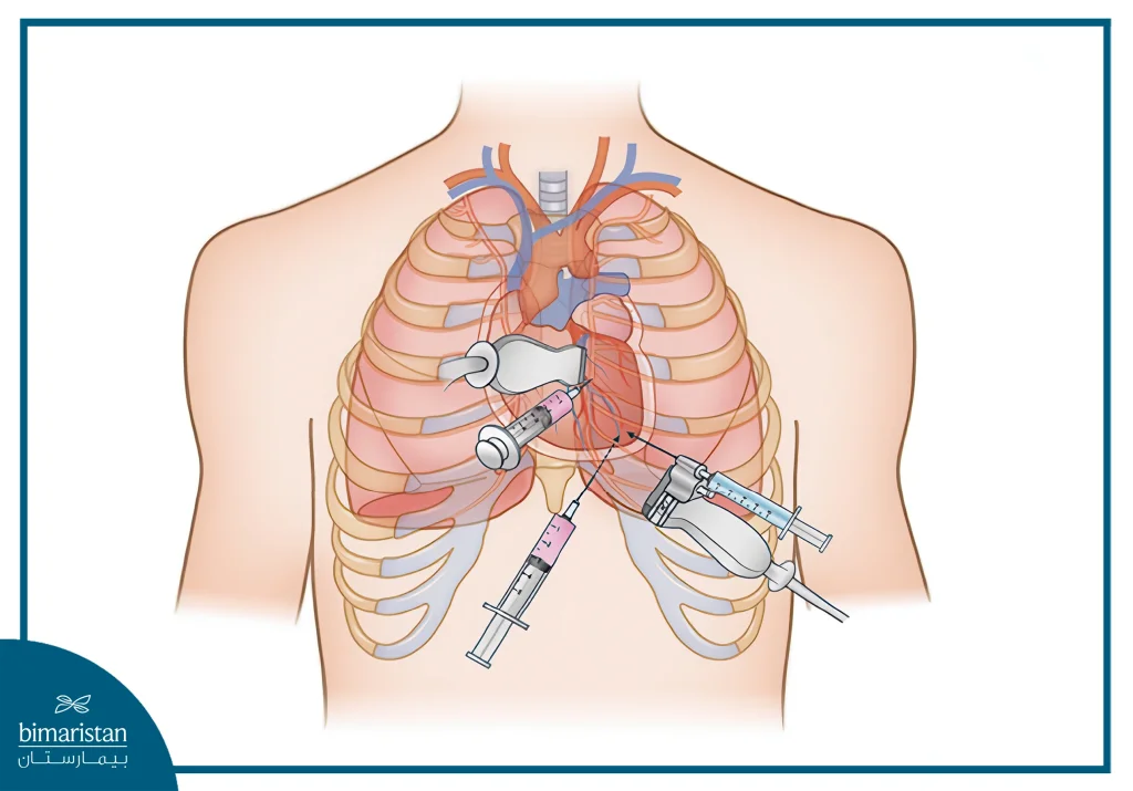
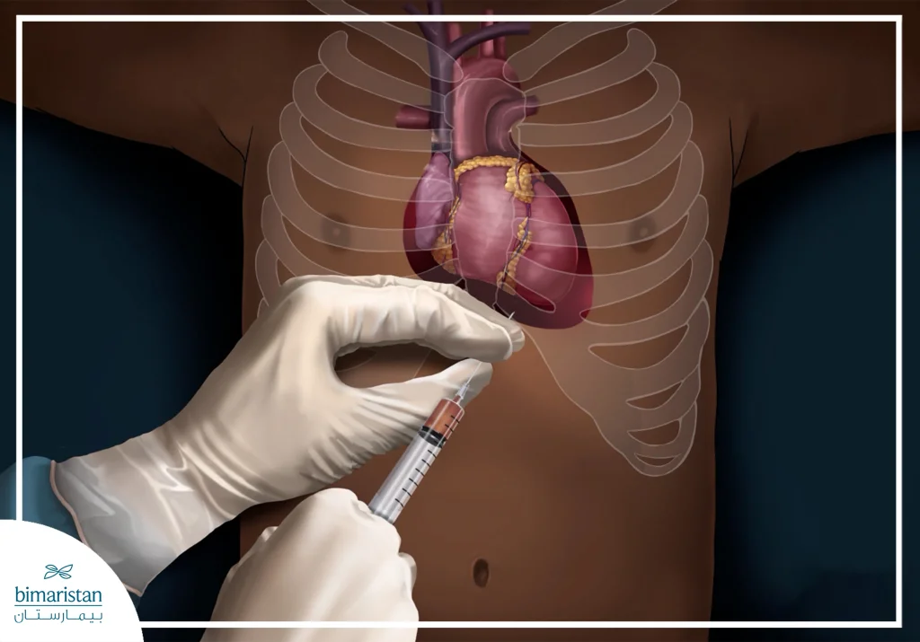
It is an emergency that needs immediate attention to treat a pericardial effusion. As a rule, the pressure here is relieved by a pericardial puncture, through which the amount of pericardial effusion is treated by fluid absorption.
How is pericardial effusion diagnosed?
Rapid diagnosis can now be made using ultrasound (echocardiography). A pericardial effusion can also be seen well on a CT scan of the heart. Additionally, the fluid in the effusion can be examined cytologically for malignant cells and bacteria following paracentesis. Analyzing the puncture fluid includes the following aspects:
- Macroscopic assessment: Such as blood, secretions, and lymph
- Microbiological examination
- Molecular bioassay (such as PCR analysis for viral pericarditis)
- Determine the number of cells
- Screening for malignant cells
Laboratory tests may also provide information about the underlying cause of the pericardial effusion, for example:
- Increased CRP in the bacterial cause
- Elevated kidney marker values in uremic poisoning
- Diagnosis of antibodies in autoimmune pericarditis
How is pericardial effusion treated?
A small pericardial effusion does not require treatment.
Depending on the cause of the disease, drug therapy can be started. However, in the case of larger effusions, ambulatory treatment should be performed by pericardiocentesis, where the effusion is withdrawn through a cannula. The puncture is performed under echocardiography, allowing the doctor to maintain control and accurately determine the location and technique of the puncture.
Another option is pericardial effusion drainage. Here, the pericardial effusion is drained via a catheter. In contrast, in the case of recurrent chronic pericarditis, a pericardiotomy is performed through the skin, for example. As with a puncture, this is also performed through the chest wall below the sternum. With the help of a catheter and a balloon, pressurized air is used to blow a hole in the pericardial wall.
The main advantage of this procedure is that the pericardial effusion can initially flow over a longer period of time, and conventional treatment is possible, depending on the nature of the flow of the pericardial effusion. There is also the option of placing a window in the pericardium, where a “window” is cut into the pericardium so that the pericardial effusion can flow better, and the treatment of the pericardial effusion depends on the amount of fluid drained.
In rare and severe cases of pericardial effusion, acute or chronic, it may be necessary to remove the pericardium. Especially when the scarring is so severe for the last effusion that it severely impedes heart activity, even if it heals. For example, an armored heart develops as a result of pericarditis. Pericardial removal is also known in medicine as pericardiectomy.

Endoscopic pericardial window for pericardial effusion
A hole is made in the pericardium by thoracoscopy so that the effusion can be drained or sucked out.
What preparations are being made?
Before the operation, an attempt is generally made to relieve the effusion by making a hole in the pericardium and inserting a tube (drainage). If this is not enough or if the effusion continues to form again, a pericardial window procedure will be resorted to. The operation is performed under general anesthesia with the usual preparations for anesthesia.
How does the process work?
For pericardial openings, the thoracoscope is inserted into the chest cavity through an incision approximately 10 centimeters below the left fifth rib arch. An opening (window) is cut in the pericardium under camera guidance so that the fluid can be emptied into the chest cavity. There, it is absorbed by the pleura (pleura). After the pericardial window, a drainage tube is usually placed to facilitate drainage. The procedure takes about an hour. The tube is removed after two to three days. The entire hospital stay generally takes 4 to 5 days.
What is the success rate of the treatment?
The results are usually very good. The effusion is eliminated with a pericardial window. The “hole” in the pericardium does not affect the heart. For better results, the underlying disease that caused the effusion must be treated.

What are the complications or risks of treatment?
This is a low-risk procedure and generally does not involve any complications. However, as with all operations, infection, bleeding, or blood clots can occur in rare cases.
What happens after the procedure?
After the pericardial window, ultrasound scans are necessary the first time after the procedure to check the progress and to ensure that no new effusion has formed.
Pericardiectomy
Chronic inflammation of the pericardium can cause the pericardium to thicken over time and form a shell that constricts the heart. This phenomenon is also called an armored heart. In this case, surgical treatment consists of partial or complete removal of the pericardium (pericardiectomy).
What preparations are made before pericardiectomy?
Pericardiectomy is performed under general anesthesia. Accordingly, the usual clarifications are necessary before the operation, such as blood tests, ECG, and blood pressure measurement. You are hospitalized the day before the operation.
How is a pericardiectomy performed?
For a pericardiectomy, the chest cavity is opened through a thoracotomy, an incision between the ribs. The surgeon then wholly or partially cuts out the pericardium, depending on the extent of the pericardial obstruction. The removed pericardium does not need to be replaced. The heart can move freely in the chest cavity without the pericardium. The procedure is usually possible without the use of a heart-lung machine.
What is the success rate of pericardiectomy?
The results are excellent. After a short period of getting used to the new situation without the pericardium, the heart works properly again.
What are the complications or risks of pericardiectomy treatment?
Pericardiectomy is considered low-risk and generally without complications. However, as with all operations, infection, bleeding, or blood clots can occur in rare cases.
What happens after a pericardiectomy?
Since the heart must get used to the new situation, intensive monitoring is necessary for a few days after the procedure, and blood circulation must be supported with medication. Until the wounds are completely healed, you should avoid heavy lifting and stay away from harsh physical exertion. After the operation, heart function is checked regularly over a longer period of time.
Sources:

